- Arms
- Arms I ca. 50% of ML
- Arms III ca. 100% of ML
- Arms IV ca. 175% 0f ML
- Largest suckers globular.
- Largest arm suckers with 12-15 broad, rounded to truncate teeth, fused laterally in distal 1/2 - 2/3 of ring; proximal portion smooth; more distal suckers with thin blunt teeth; more proximal suckers with broader, rounded teeth.
 Click on an image to view larger version & data in a new windowClick on an image to view larger version & data in a new window
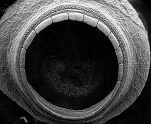
Figure. Oral view of arm sucker 14, arm III, of C. spoeli, 75 mm ML, central North Atlantic, off Bermuda. Scanning electron micrograph by R. Young.
Comparisons of arm sucker dentition from different geographic areas can be seen here
- Tentacles
- Club length 65% of ML.
- Suckers with 7-8 long, triangular, sharply-pointed teeth over distal half of ring; no enlarged central tooth.
Click on an image to view larger version & data in a new windowClick on an image to view larger version & data in a new window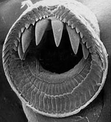
Figure. Oral view of club sucker of C. spoeli, central North Atlantic, off Bermuda, 75 mm ML. Scanning electron micrograph by R. Young.
Comparisons of club sucker dentition from different geographic areas can be seen here.
- Sucker stalks in two distinct parts (long, cyclindrical base and short, constricted portion that attaches to sucker); stalks of lateral and medial suckers about equal in length.
Click on an image to view larger version & data in a new windowClick on an image to view larger version & data in a new window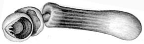
Figure. Proximal view of club sucker and stalk of C. spoeli. Note division of stalk and poorly demarcated stalk-pleating.
- Protective membranes
- Membranes in three distinctive sets with intermediate set broadest.
- Proximal set with 20-31 slender, separate trabeculae (ca. 25-35% of club length).
- Intermediate set without distinct trabeculae (forms fleshy membrane) but opposite 13-15 rows of suckers (about 25-30% of CL).
- Distal set with slender, separate, trabeculae opposite 30-31 rows of suckers (about 35-45% of CL).
Click on an image to view larger version & data in a new windowClick on an image to view larger version & data in a new window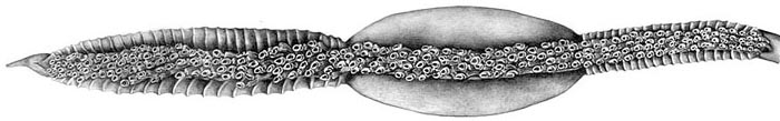
Figure. Oral view of tentacular club of C. spoeli. Note three-part division of protective membranes.
Click on an image to view larger version & data in a new window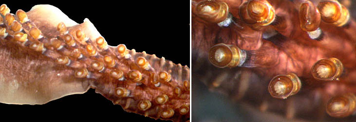
Figure. Tentacular club of C. spoeli, Hawaiian waters, preserved. Left - Oral view of part of the proximal and middle portions of the club. Right - Closer view of the proximal portion; note the pleated skirt on the sucker stalk. Photographs by R. Young.
- Head
- Head length ca. 40% of ML.
- Funnel
- Funnel-locking apparatus with broad, protruding tragus and broad, well-developed antitragus; both overhang.
Click on an image to view larger version & data in a new windowClick on an image to view larger version & data in a new window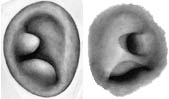
Figure. Funnel-mantle locking apparatus of C. spoeli. Left - Ventral view of funnel component. Right - Dorsal view of mantle component.
- Funnel-locking apparatus with broad, protruding tragus and broad, well-developed antitragus; both overhang.
- Fins
- Fin length 40-45% of ML.
- Fin width 40-45% of ML.
- Photophores
- Eyeball - Two series: lateral series, 8 organs = 1+6+1 or 7 organs = 1+5+1 or 6 organs = 1+4+1; medial series, seven organs = 1+5+1 or six organs = 1+4+1.
Click on an image to view larger version & data in a new windowClick on an image to view larger version & data in a new window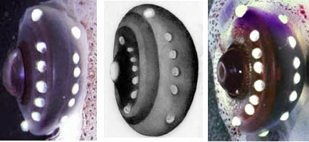
Figure. Ventral view of eyeball of C. spoeli showing photophore arrangement. Note variation in photophore numbers in these three eyes from left to right: 1+5+1:1+1+4+1 (Hawaii); 1+5+1:1+4+1 (Atlantic); 1+6+1:1+5+1 (Hawaii). Photographs by R. Young, drawing by J. R. Schroeder.
- Viscera: two, large photophores.
- Club-tip photophore small, with long papilla.
Click on an image to view larger version & data in a new windowClick on an image to view larger version & data in a new window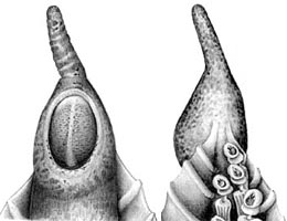
Figure. Club-tip photophore of C. spoeli. Left - Aboral view Note large terminal papilla and discontinuity (light band) down center of photophore. Right - Oral view and distal club suckers. Drawing by J. R. Schroeder.
- One photophore embedded in aboral surface of club at proximal end opposite trabecula no. 4 (counting from proximal, very small trabecula).
- Eyeball - Two series: lateral series, 8 organs = 1+6+1 or 7 organs = 1+5+1 or 6 organs = 1+4+1; medial series, seven organs = 1+5+1 or six organs = 1+4+1.
- Pigmentation
- Club-tip photophore with chromatophores only.
- Club pigment in epithelial cells rather than chromatophore organs except at distal tip.
- Sucker stalks pigmented and with long, poorly demarcated pleats.
- Olfactory organ with chromatophores on stalk and bulb.
- Buccal membrane unpigmented.
Click on an image to view larger version & data in a new windowClick on an image to view larger version & data in a new window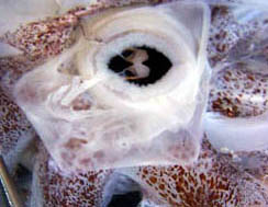
Figure. Oral view of buccal region of C. spoeli showing unpigmented buccal membrane.
Chiroteuthis spoeli: Description continued
Richard E. Young and Clyde F. E. RoperAbout This Page

University of Hawaii, Honolulu, HI, USA
Smithsonian Institution, Washington, D. C., USA
Page copyright © 1999 and
 Page: Tree of Life
Chiroteuthis spoeli: Description continued
Authored by
Richard E. Young and Clyde F. E. Roper.
The TEXT of this page is licensed under the
Creative Commons Attribution-NonCommercial License - Version 3.0. Note that images and other media
featured on this page are each governed by their own license, and they may or may not be available
for reuse. Click on an image or a media link to access the media data window, which provides the
relevant licensing information. For the general terms and conditions of ToL material reuse and
redistribution, please see the Tree of Life Copyright
Policies.
Page: Tree of Life
Chiroteuthis spoeli: Description continued
Authored by
Richard E. Young and Clyde F. E. Roper.
The TEXT of this page is licensed under the
Creative Commons Attribution-NonCommercial License - Version 3.0. Note that images and other media
featured on this page are each governed by their own license, and they may or may not be available
for reuse. Click on an image or a media link to access the media data window, which provides the
relevant licensing information. For the general terms and conditions of ToL material reuse and
redistribution, please see the Tree of Life Copyright
Policies.


