I. External structure

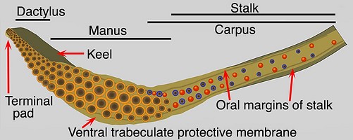
Figure. Tentacular club of T. rhombus, oral view. Blue dots - Carpal suckers. Red dots - Carpal knobs. Drawing by R. Young.
Features:
- Carpus: Present. Two carpal suckers and knobs along dorsal margin of manus; two series of alternating suckers and knobs along distal half of tentacular stalk. These two parts of carpus separated by small gap.
- Club divisions: Carpus, manus, dactylus and terminal pad.
- Club shape: Expanded manus with narrow dactylus generally having a dorsal curvature.
- Sucker series: Four, regular, longitudinal series except for few suckers at proximal end of club.
- Trabeculate protective membranes: Trabeculae (one per lateral sucker) present (can be difficult to distinguish); dorsal membrane low to virtually absent on distal half of dactylus; dorsal and ventral membranes very broadly separated at proximal end of club.
- Oral margins of stalk: Present; muscular but lack distinct trabeculae; located at lateral edges of stalk; continuous with trabeculate protective membranes of stalk; dorsal margin extends length of carpus; ventral margin extends much further.
- Aboral margin of stalk: Present and very prominent; positioned on dorsal-aboral corner of stalk; extends along much (all ?) of tentacular stalk and onto club; terminates just distal to beginning of dactylus (overlaps Keel but offset from it dorsally); apparently muscular but with large fluid-filled spaces.
- Keel: Extends to midlevel of manus; positioned dorsally throughout its length.
- Terminal pad: Present, distinct.
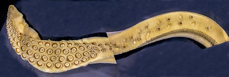
Figure. T. rhombus. Oral view of a club and stalk that was twisted but photographed at two different angles then artificially combined.
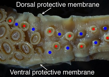
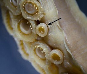
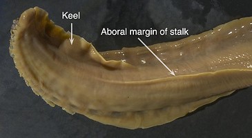
Figure. T. rhombus, different specimen. Top left - Carpal locking apparatus (red dots - carpal suckers, blue dots - carpal knobs). Note the continuation of the protective membrane from the manus to the oral margins of the stalk. Top right - Photomicrograph of the distal-most carpal sucker (arrow) showing the presence of teeth. Typically all carpal suckers have smooth inner rings. Here is an exception. Bottom, left - Aboral view of most of the club showing the overlapping but offset positions of the keel and aboral margin of the stalk. Bottom, right - Oral view of the Terminal pad stained with methylene blue stain. Yellow dots indicate terminal suckers of dactylus with toothed inner sucker rings; the remaining suckers are smooth-ringed and belong to the terminal pad. Note that the largest and most proximal of the latter looks like it should belong to the carpus.
II. External structure: Oral surface manus without suckers
- Musculature between suckers, suckers and trabeculae, and trabeculae complex and poorly understood. There are no "open canals" or deep pockets surrounding the medial sucker-basess. Seawater does penetrate in restricted pockets especially between the bases of trabeculae and the lateral bases of the medial sucker-stalks (yellow arrows in image below).
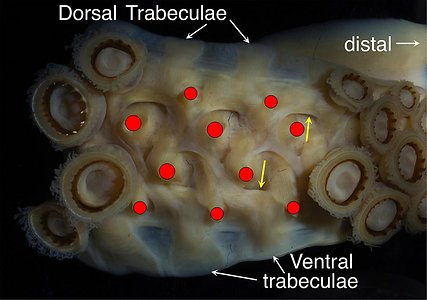
Figure. T. rhombus. Oral view of the distal manus with some suckers removed showing the complex arrangement of surface muscles. Note the involvement of the broad trabeculae of both dorsal and ventral membranes in the musculature. Red dots - Mark where sucker stalks were cut. Yellow arrows - Indicates pockets between bases of trabeculae their associated bases of medial suckers that allows seawater penetration (see next section). Note that the pockets have a more fully formed openings in the dorsal set.
III. Internal structure: Cross-section of the manus
- Organization: Manus somewhat bilaterally symmetrical with stalks to lateral and medial suckers about the same length (sucker sizes similar). Oral surface of manus very flat when in relaxed state.
- Trabeculate protective membranes: Dorsal and ventral membranes large and equally developed.
- Core shape: Flattened oral and lateral sides.
- Keel: Not present in sections (i.e., keel ended distal to sections). Weakly-muscular aboral margin of stalk present and located slightly ventral of mid-aboral line.
- Attachment of sucker stalks and trabeculae: Sucker stalks attach mostly to club core with some attachment to other sucker stalks; trabeculae attach to both the sucker stalks and club core. The complex musculature of the club is poorly understood.
- Open canals: Absent. Each trabecula is undercut by a water pocket that overrides lateral base of medial sucker-stalk (yellow arrow in previous image). Click on an image to view larger version & data in a new window
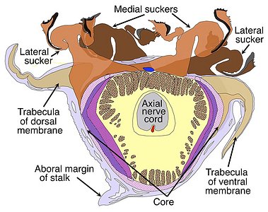
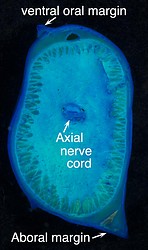
Figure. T. rhombus. Left - Male, 144 mm ML. View from more distal side of cut segment. Cross-section through the manus showing the basic structure of the club core, and the attachment of the keel, sucker stalks and trabeculae of the protective membrane. Most of the core outside the axial nerve cord is composed of muscles. Dots on drawing areas indicate primarily longitudinal muscles. Other colors and fill-lines indicate more complex musculature. The red dot on the aboral side of the nerve cord is the major artery of the club; the blue dot near the central of the oral surface is the major vein of the club. The drawing, by R. Young, is a compilation taken from histological cross-sections. Note the water pocket beneath the trabecula of the dorsal membrane. Right - Cross-sectional cut with a scalpel of the tentacular stalk just proximal of the club, for comparison with the club.
II. Internal structure: Transverse cut through the mid-dactylus (no histological sections available)
- Organization: Strong asymmetry with very long stalks for the ventrolateral suckers grading to small stalks for dorsolateral suckers. Oral surface of dactylus basically flat, when in relaxed state, and in same plane with that of manus.
- Trabeculate protective membranes: Ventral trabecula and membrane well developed; dorsal counterparts greatly reduced.
- Core shape: Oral side (as seen in manus) flattened but tilted ventrally.
- Keel: Large; attached to dorsal side of core; oral surface (when relaxed) probably parallel to that of combined suckers.
- Attachment of sucker stalks and trabeculae: Uncertain.
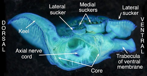
Figure. T. rhombus, transverse cut through the mid-dactylus (only 3 suckers clearly visible due to the oblique arrangement of transverse sucker rows) showing the basic structure. Cut stained with methylene blue stain. Note that no trace of a dorsal protective membrane is apparent.


