Drawings below are first made from photographs. The drawings should then be compared, under a microscope, with the preserved squid from which the photographs were made. This greatly reduces the errors that can occur in interpreting the photophore distributions. Use caution, therefore, on this page in viewing the color and distribution of the dots (representing photophores) as their distributions have not yet been confirmed by comparing the drawings against the actual specimen under the microscope.

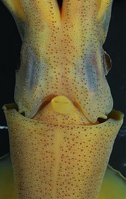
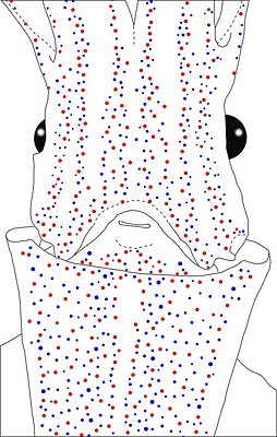
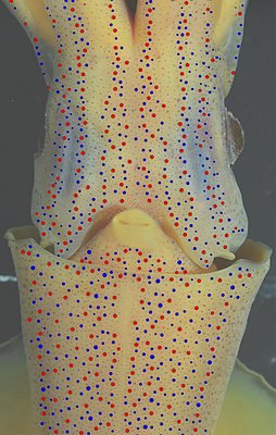
Figure. Ventral views of the integumental photophores. Left - Photograph of the preserved squid. Middle - Outline drawing from photograph with all integumental photophores represented by colored dots. Right - Photograph with colored dots superimposed on integumental photophores. Red dots - Complex photophores. Blue dots - Non-complex photophores. Images by R. Young.
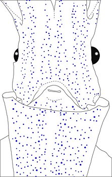
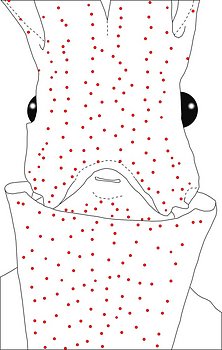
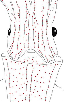
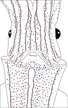
Figure. Same squid, same view as above. Left - Drawing showing only the non-complex (blue) photophores. Left middle - Drawing showing only the complex (red) photophores. Right middle - Same as previous drawing with lines connecting red photophores to aid comparisons of photophore patterns between species. Right - Drawing showing all photophores and lines showing red photophore patterns. Images by R. Young.
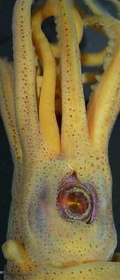
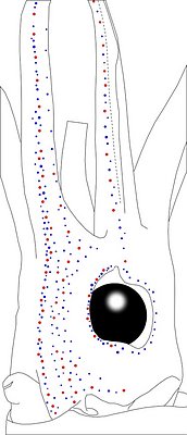
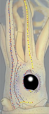
Figure. Side and slightly oblique view of the head of the same squid (29 mm ML) as above. Left - Photograph of the preserved squid. Middle - Outline drawing from photograph with all integumental photophores represented by colored dots. Right - Outline drawing over dimmed photograph with all integumental photophores represented by colored dots, and with lines connecting some photophores. Red dots - Complex photophores. Blue dots - Non-complex photophores. Lines connecting photophores assist in understanding the organization of the photophores. Black lines - Patterns based on red photophores. Yellow line - Pattern based on red and blue photophores. Images by R. Young.
Abraliopsis sp. Z exhibits and unusual amount of variability in the arrangement of ventral head photophores. The most common pattern is the one illustrated above. One of the more extreme variations, similar to a fully scattered arrangement, is shown below.
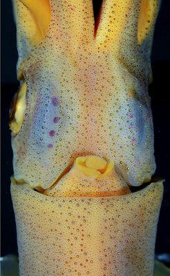
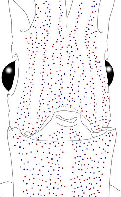
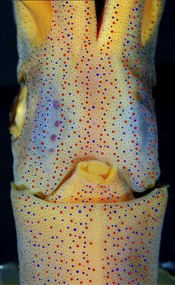
Figure. Ventral views of the integumental photophores. Left - Photograph of the preserved squid. Middle - Outline drawing from photograph with all integumental photophores represented by colored dots. Right - Photograph with colored dots superimposed on integumental photophores. Red dots - Complex photophores. Blue dots - Non-complex photophores. Images by R. Young.
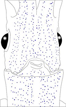
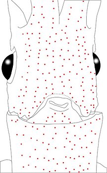
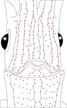
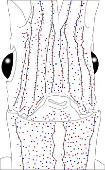
Figure. Same view, same squid (35 mm ML) as above. Left - Drawing showing only the non-complex (blue) photophores. Left middle - Drawing showing only the complex (red) photophores. Right middle - Same drawing with lines connecting red photophores to aid comparisons of photophore patterns between species. Right - Drawing showing all photophores; lines connecting red dots aid in understanding the arrangement of the photophores. Images by R. Young.
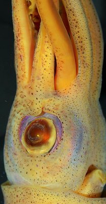
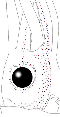
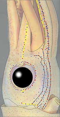
Figure. Oblique view of the head and proximal regions of the arms of the same squid (35 mm ML) as above. Left - Photograph of the preserved squid. Middle - Outline drawing from photograph with all integumental photophores represented by colored dots. Right - Outline drawing over dimmed photograph with all integumental photophores represented by colored dots, and with lines connecting some photophores. Red dots - Complex photophores. Blue dots - Non-complex photophores. Lines connecting photophores assist in understanding the organization of the photophores. Black lines - Patterns based on red photophores. Yellow line - Pattern based on red and blue photophores. Images by R. Young.
Figure. Ventral views of the mantles of A. sp. Z. Top - Female, 29 mm ML. Bottom - Female, 35 mm ML. Note the well-defined, bare midline stripe of the Median Mantle Sector that extends the full length of the mantle in both squid. Photographs by R. Young.
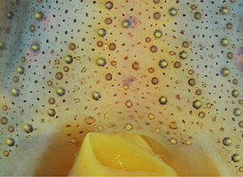
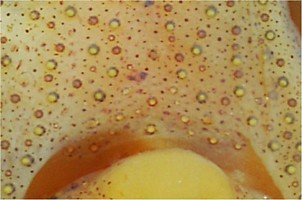
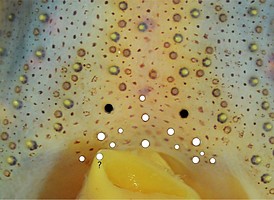
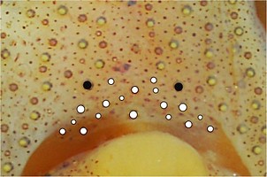
Table. Ventral view of the funnel-groove region of Abraliopsis sp. Z. Top tier - Funnel groove showing photophores of funnel groove and surrounding tissue. Middle tier - In the photograph, white dots placed over the "white" photophores of the funnel groove. Black dots placed over the most posteromedial "red" photophores of the ventral head to mark the anterolateral edges of the funnel groove. Bottom tier - Same pattern of dots as in photographs. Colors, lines - Aid in comparison of patterns between species. Open dots - Represent photophores that are either of uncertain status as "white" photophores or have variable presence. Images by R. Young. Comparison of white photophore patterns for all Abraliopsis species can be found here.
| Mature male, 28 mm ML | Mature female, 35 mm ML |
|---|---|
 |  |
 |  |
 |  |
Comments: Summary of photophore distributions. (photophore definitions found here)
ARMS:
Medial Arm IV Series - Continuous; reaches 75-80% of arm length.
Central Arm IV Sector - No photophores other than those adjacent to arm IV Series.
Lateral Arm IV Series - Continuous; extends to arm tip.
Lateral Membrane Series of arm IV - Continuous, reaches at least 2/3 of arm length.
Arm III Series - Continuous; eaches nearly 90% of arm length.
HEAD - Typical scattered pattern with:
Median Head Series - Absent.
Median Head Sector - Few photophores. Noticeably photophore-poor in center line.
Medial Head Series - Medial Head Series "A" + Medial Head Series "B" + Head Sector "AB" (This sector contained between MHS "A" and MHS "B." These 2 series and containing sector defined only for Abraliopsis sp. Z) generally contain most photophores medial to Lateral Head Series.
Lateral Head Sector - (For this species: this sector lies between Medial Head Series "B" and Lateral Head Series.) generally a photophore-poor region.
Window Sector - Few scattered blues at either end plus two reds in Olfactory Series (at 29 mm ML); otherwise without photophores.
First Lateral Window Series - Continuous series with at least 8 red photophores at 29 mm ML.
Second Lateral Window Series - Discontinuous series at 29 mm ML with 2 segments, posterior segment with 2 reds, anterior segment with 4 reds. Continuous series at 35 mm ML with at least 9 red photphores.
Eyelid Series - Red photophores only on ventral half of eyelid. Posterior Eyelid Series with one large blue photophores displaced slightly posteriorly from eyelid series. Formula: RbBB.
Occipital Series - RbRbR at 28 mm ML; RbRbbbR at 29 mm ML; RbRbbR at 35 mm ML.
Lateral Funnel-groove Series - Two segments not well aligned; posterior segment with 3 reds, anterior segment with 3 reds at 29 and 35 mm ML.
Olfactory Series - 2 red photophores at 29 and 35 mm ML. Posterior member just posteroventral to first (= most posterior) photophore of First Lateral Window Series and nearest red photophore to olfactory organ on 2nd occipital fold. Anterior member anteroventrally to posterior member. 1 red photophore and one blue (anterior member) at 28 mm ML.
White Patch (funnel groove) - Moderate pattern (red, green, blue + yellow with size).
FUNNEL: Patches not well demarcated.
Medial Patch - Scattered red photophores.
Lateral Patch - Two red photophores (in young at least - see photo on previous page).
MANTLE - Typical scattered pattern with:
Median Mantle Sector - Well defined in anterior 15-20% of the ML with virtually no blue photophores medial to either Medial Mantle Series. Bare region of Medial Mantle Sector extends full length of mantle.
Medial Mantle Series - Irregular.
Mediolateral Mantle Sector - Scattered photophores; Lateral Mantle Series not apparent.
Mantle-angle Series - Recognizable.
Comments 2
Abraliopsis sp. Z is most easily recognized by the wide, bare strip of the Median Mantle Sector that extends virtually to the anterior mantle margin (This feature is presumably shared with A. tui). It also has an unusually large number of photophores. The ventral head photophores tend to show a pattern with relatively few photophores around the median line and few in a modified Lateral Head Sector, leaving most photophores in a swath between these two areas. This swath of photophores recalls the complex Lateral Head Series of A. gilchristi. Unfortunately the pattern on the head of A. sp. Z is variable enough that this pattern is not always apparent.


