
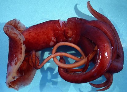
Figure Ventral view of the tropical form of M. agassizii, 90 mm ML, Gulf of Mexico. Collected by T. Sutton, HBOI. Photograph by R. Young.
- Arms
- Arm formula: IV>II>III>I.
- Arm II length ca. 90% arm II length.
- Arm II ca. 50-60% 0f ML.
- Arms with low protective membranes.
- Arm suckers with ca. 12-15 pointed teeth on distal margins of inner rings.
- Largest arm suckers of arms III and IV equal in size; ca. 0.6 mm across outer ring in 79 mm ML squid.
Click on an image to view larger version & data in a new window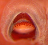
Figure. Oral-oblique view of an arm sucker of the tropical form of M. agassizii, 90 mm ML. Photograph by R. Young
- Tentacles
- Suckers on club smaller and more widely separated proximally (typical of the genus). Sucker diameter ca. 0.16 mm in midclub, ca. 0.10 at base in 79 mm ML squid.
- Sucker-bearing club completely encircles tentacle in terminal 10-15% of club; Purple line marke the remnant of the aboral surface and can be followed to club tip.
- Club covers over 50 of tentacle circumference at its abruptly terminating base.
- Head
- Rounded but distinct occipital crest without occipital folds.
- Funnel pocket present.
- Stalk of olfactory organ more slender than sensory bulb and weakly pigmented.
Click on an image to view larger version & data in a new windowClick on an image to view larger version & data in a new window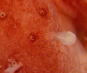
Figure. Ventral view of the olfactory organ of the tropical form of M. agassizii, 90 mm ML. Photograph by R. Young. Collected by T. Sutton, HBOI
- Fins
- Fins together nearly circular in outline.
- Tubercules
- Integumental tubercules absent.
- Integumental tubercules absent.
- Photophores and pigmentation
- Integument with typical integumental photophores, ringed chromatophores and white sphere (see photograph of funnel organ above).
- Most integumental chromatophores ringed and well separated.
- Eyelid photophore present, small (ca 0.2 mm in 79 mm ML squid), with distinct window.
- Arms IV with two, somewhat irregular, series of photophores; distally becoming a single series and breaking into separate patches.
- Arms III with single series of photophores to arm tip on aboral-ventral margin.
Click on an image to view larger version & data in a new window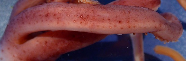
Figure. Ventral view of a midregion of arm IV of the tropical form of M. agassizii, 79 mm ML. Photograph by R. Young.
Click on an image to view larger version & data in a new window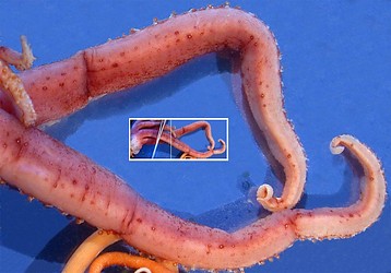
Figure. Ventral view of the distal portion (distal to the vertical white line in the insert) of arms IV of the tropical form of M. agassizii, 79 mm ML. The background has been cleaned-up in Photoshop. Photograph by R. Young.
- Photophores on funnel and mantle scattered.
- Few small photophores on midregion of cyclindrical, non-muscular mantle extension between fins but patch of large photophores just proximal to tail.
Click on an image to view larger version & data in a new window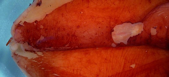
Figure. Ventral view of photophores on non-muscular, posterior extension of the mantle of the tropical form of M. agassizii. Photograph by R. Young. Collected by T. Sutton, HBOI
- Most pigmentation not in chromatophores.
- Inner mantle wall unpigmented; visceral sac with light pigmentation posteriorly.
Click on an image to view larger version & data in a new window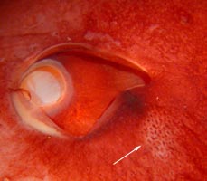
Figure. Lateral view of eyelid region of the tropical form of M. agassizii, 90 mm ML, preserved. Eyelid photophore is covered laterally by dark pigment but the window through which the bioluminescence passes is apparent (arrow) and has a covering of small photophores. Photograph by R. Young.
Click on an image to view larger version & data in a new window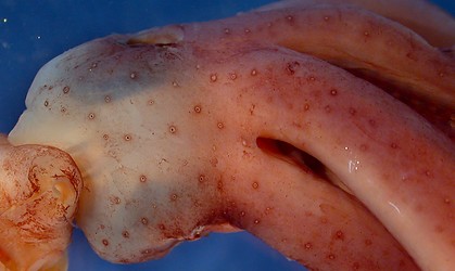
Figure. Ventral view of the head and bases of arms IV of the tropical form of M. agassizii, 79 mm ML, showing scattered pattern of large and small photophores. The skin was slightly abraided on this specimen making the pattern of the deep photophores apparent. Photograph by R. Young.
- Measurements
*Where possible, right and left measurements averaged.Squid SC 98 21 SC 95 T8 Sex Imm. female Imm. male Mantle length 90 79 Tail lenght
missing missing Fin length 60 48 Fin width 66 51 Head width 26 20 Eye diameter 15 10.5 Arm I, length: Left / Right 43.5 29.5 Arm II, length 58.5 43.5 Arm III, length 52.5 38.5 Arm IV, length 121.5 92.5 Tentacle length 189 205 Club length (% of tentacle length) 65.5 62% Largest club sucker diam. 0.16
Largest arm sucker diam.
0.6 across ring
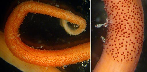
Figure. Tentacular club of the tropical form of M. agassizii. Left - Aboral view of club tip showing club completely encircling tentacle, 79 mm ML, Gulf of Mexico. Right - Oral view of club base showing abrubt termination, 90 mm ML, Gulf of Mexico. Photographs by R. Young.


