|
ID
|
Thumbnail |
Media Data |
| 713 |
|
|
Scientific Name
|
Astraspis desiderata
|
|
Location
|
Colorado
|
|
Comments
|
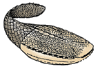
Astraspids are still poorly known but recent discoveries of partially complete specimens of Astraspis desiderata, from the Ordovician of Colorado, have considerably increased their knowledge. Their dorsal headshield is made up by large, polygonal bone units and the gill openings are situated more dorsally than in arandaspids.
|
|
Reference
|
After Janvier 1996, modified from Elliott, D. K. (1987). A reassessment of Astraspis desiderata, the oldest North American vertebrate. Science, 237:190-192.
|
|
Specimen Condition
|
Fossil -- Period: Ordovician
|
|
Image Use
|
 This media file is licensed under the Creative Commons Attribution License - Version 3.0. This media file is licensed under the Creative Commons Attribution License - Version 3.0.
|
|
Copyright
|
© 1997

|
|
Attached to Group
|
Astraspida: view page image collection
|
|
Title
|
astraspida.gif
|
|
Image Type
|
Drawing/Painting
|
|
Image Content
|
Specimen(s)
|
|
ID
|
713
|
|
| 722 |
|
|
Scientific Name
|
Tauraspis, Hoelaspis, Tremataspis, Zenaspis
|
|
Comments
|
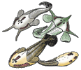
Osteostracans are known from the Silurian and Devonian of Europe, Siberia and North America. They are characterized by peculiar "cephalic fields" of unknown function, on the dorsal surface of the head-shield (red). Although jawless, they share with jawed vertebrates well-developed paired fins, an epicercal tail, cellular bone, and a sclerotic ring in eyes. Their mouth and gill opening are ventrally placed, as in galeaspids and pituriaspids. Their median, dorsal, nasohypophysial aperture, anterior to the eyes, is strikingly similar to that of lampreys but is now regarded as a convergence. All the osteostracans reconstructed here belong to the major clade Cornuata, whose generalized morphology is exemplified by the zenaspidid Zenaspis (bottom left). Some highly derived head-shield morphologies are exemplified by the benneviaspidids Hoelaspis (top right) and Tauraspis (top left), or the thyestiid Tremataspis (bottom right). The latter has lost the paired fins, possibly as a consequence of an adaptation to burrowing habits.
|
|
Reference
|
based on Janvier, P. 1985. Les Céphalaspides du Spitsberg. Anatomie, phylogénie et systématique des Ostéostracés siluro-dévoniens. Révision des Ostéostracés de la Formation de Wood Bay (Dévonien inférieur du Spitsberg). Cahiers de Paléontologie, Centre national de la Recherche scientifique, Paris. AND Janvier, P. 1985. Les Thyestidiens (Osteostraci) du Silurien de Saaremaa (Estonie). Première partie: Morphologie et anatomie, Annales de Paléontologie, 71(2), p.83-147. Deuxième partie: Analyse phylogénétique, répartition stratigraphique, remarques sur les genres Auchenaspis, Timanaspis, Tyriaspis, Didymaspis, Sclerodus et Tannuaspis. Annales de Paléontologie, 71(3), p.187-216. AND Mark-Kurik, E. & Janvier, P. 1995. Early Devonian osteostracans from Severnaya Zemlya, Russia. Journal of Vertebrate Paleontology. 15(3):449-462.
|
|
Image Use
|
 This media file is licensed under the Creative Commons Attribution License - Version 3.0. This media file is licensed under the Creative Commons Attribution License - Version 3.0.
|
|
Copyright
|
© 1997

|
|
Attached to Group
|
Osteostraci: view page image collection
|
|
Title
|
osteostraci.gif
|
|
Image Type
|
Drawing/Painting
|
|
Image Content
|
Specimen(s)
|
|
ID
|
722
|
|
| 1056 |
|
|
Comments
|
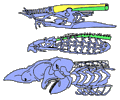
The craniates are characterized by a skull; that is, a complex ensemble of skeletal elements which surrounds the brain and sensory capsules. The skull of hagfishes (top) consists of cartilaginous bars (blue), but the brain is mostly surrounded by a fibrous sheath (yellow) underlain by the notochord (green). The skull of lampreys (middle) has a more elaborate braincase and comprises a large "branchial basket" surrounding the gills. In the gnathostomes (bottom), the braincase is generally closed (after Janvier 1996b).
|
|
Reference
|
after Janvier, P. (1996). Early Vertebrates. Oxford Monographs in Geology and Geophysics, 33, Oxford University Press, Oxford.
|
|
Body Part
|
skull
|
|
Image Use
|
 This media file is licensed under the Creative Commons Attribution License - Version 3.0. This media file is licensed under the Creative Commons Attribution License - Version 3.0.
|
|
Copyright
|
©

|
|
Attached to Group
|
Craniata: view page image collection
|
|
Title
|
craniata.gif
|
|
Image Type
|
Drawing/Painting
|
|
Image Content
|
Body Parts
|
|
ID
|
1056
|
|
| 1808 |
|
|
Comments
|
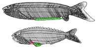
Anaspids are characterized by a large, tri-radiate spine (red) posteriorly to the series of branchial openings. Typical anaspids are restricted to the Silurian but some doubtful forms occur in the Late Devonian. It is assumed that the most primitive anaspids, such as Pharyngolepis (top), possessed a long, ribon-shaped, ventrolateral fin-fold (green). More advanced forms, such as Rhyncholepis (bottom), possessed a shorter paired fin-fold (green) and enlarged, spine-shaped, median dorsal scutes.
|
|
Reference
|
after Ritchie, A. (1964). New light on the morphology of the Norwegian Anaspida. Skrifter utgitt av det Norske Videnskaps-Akademi, 1, Matematisk-Naturvidenskapslige Klasse, 14, 1-35.
and Ritchie, A. (1980). The Late Silurian anaspid genus Rhyncholepis from Oesel, Estonia, and Ringerike, Norway. American Museum Novitates, 2699, 1-18.
|
|
Specimen Condition
|
Fossil
|
|
Image Use
|
 This media file is licensed under the Creative Commons Attribution License - Version 3.0. This media file is licensed under the Creative Commons Attribution License - Version 3.0.
|
|
Copyright
|
© 1997

|
|
Attached to Group
|
Anaspida: view page image collection
|
|
Title
|
anaspida.gif
|
|
Image Type
|
Drawing/Painting
|
|
Image Content
|
Specimen(s)
|
|
ID
|
1808
|
|
| 1895 |
|
|
Scientific Name
|
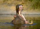
Hippopotamus amphibius
|
|
Location
|
Okavango Delta of Botswana
|
|
Sex
|
Male
|
|
Body Part
|
head
|
|
Copyright
|
© 1997 Greg and Marybeth Dimijian

|
|
Image Use
|
ToL use only
|
|
Attached to Group
|
Hippopotamus amphibius (Hippopotamidae): view page image collection
|
|
Title
|
01035hippo.jpg
|
|
Image Type
|
Photograph
|
|
Image Content
|
Specimen(s)
|
|
ALT Text
|
Hippopotamus with mouth wide open
|
|
ID
|
1895
|
|
| 2241 |

|
|
| 2346 |

|
|
Scientific Name
|
Homo sapiens
|
|
Acknowledgements
|
The Digital Human Osteology Guide
|
|
Body Part
|
skull
|
|
Copyright
|
© 1997 John Kappelman
|
|
Image Use
|
restricted
|
|
Attached to Group
|
Craniata: view page image collection
|
|
Title
|
skull.250.gif
|
|
Image Type
|
Photograph
|
|
Image Content
|
Specimen(s)
|
|
ID
|
2346
|
|
| 2367 |
|
|
Scientific Name
|
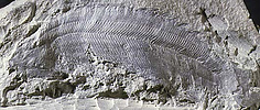
Pterygolepis nitidus
|
|
Location
|
Norway
|
|
Specimen Condition
|
Fossil -- Period: Lower Silurian
|
|
Image Use
|
 This media file is licensed under the Creative Commons Attribution License - Version 3.0. This media file is licensed under the Creative Commons Attribution License - Version 3.0.
|
|
Copyright
|
© 1997

|
|
Attached to Group
|
Pterygolepis (Vertebrata): view page image collection
|
|
Title
|
pterygolepis.jpeg
|
|
Image Type
|
Photograph
|
|
Image Content
|
Specimen(s)
|
|
ALT Text
|
Pterygolepis nitidus fossil
|
|
ID
|
2367
|
|
| 2387 |
|
|
Comments
|
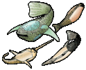
Heterostracans are the most diverse group of pteraspidomorphs and lived during the Silurian and Devonian periods. Among the most primitive heterostracans are tolypelepids (top right). Most heterostracans are pteraspidiforms, such as the pteraspidids (bottom right), protopteraspidids (bottom left) and the huge psammosteids (top left), which are the youngest known members of the group.
|
|
Reference
|
After Janvier 1996 AND Soehn, K. L. and Wilson, M. K. V. (1990). A complete, articulated heterostracan from Wenlockian (Silurian) beds of the Delorme Group, Mackenzie Mountains, Northwest territories, Canada. Journal of Vertebrate Paleontology, 10:405-419.
|
|
Specimen Condition
|
Fossil
|
|
Image Use
|
 This media file is licensed under the Creative Commons Attribution License - Version 3.0. This media file is licensed under the Creative Commons Attribution License - Version 3.0.
|
|
Copyright
|
© 1997

|
|
Attached to Group
|
Protopteraspididae (Pteraspidomorphi): view page image collection
Pteraspididae (Pteraspidomorphi): view page image collection
Tolypelepidida (Heterostraci): view page image collection
Psammosteidae (Pteraspidomorphi): view page image collection
|
|
Title
|
heterostraci.gif
|
|
Image Type
|
Drawing/Painting
|
|
Image Content
|
Specimen(s)
|
|
ID
|
2387
|
|
| 2486 |

|
|
Comments
|
The vertebrates are characterized by a vertebral column; that is, a variable number of endoskeletal elements aligned along the notochord (green) and flanking the spinal cord (yellow). In lampreys (top), the vertebral elements are only the basidorsal (red) and the interdorsals (blue). In the gnathostomes, there are in addition ventral elements, the basiventrals (purple) and interventrals (orange), and the notochord may calcify into centra (pink).
|
|
Reference
|
After Janvier, P. (1996). Early vertebrates. Oxford Monographs in Geology and Geophysics, 33, Oxford University Press, Oxford.
|
|
Body Part
|
vertebral column
|
|
Image Use
|
 This media file is licensed under the Creative Commons Attribution License - Version 3.0. This media file is licensed under the Creative Commons Attribution License - Version 3.0.
|
|
Copyright
|
©

|
|
Attached to Group
|
Vertebrata: view page image collection
|
|
Title
|
vertebrata.gif
|
|
Image Type
|
Drawing/Painting
|
|
Image Content
|
Body Parts
|
|
ID
|
2486
|
|


 This media file is licensed under the
 Go to quick links
Go to quick search
Go to navigation for this section of the ToL site
Go to detailed links for the ToL site